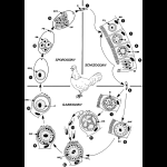
Fig. 1. Life cycle of
Eimeria spp. in chicken. For species and other hosts see
Table 1.
1 After oral uptake of sporulated oocysts the sporozoites hatch in the small intestine from the sporocysts.
2–6 After penetration, multinucleate
schizonts are formed (3) inside a
parasitophorous vacuole (
PV). The schizonts produce motile
merozoites (
DM, M), which may initiate another generation of schizonts in other intestinal cells (
2–5) or become gamonts of different sex (
7, 8).
7 Formation of multinucleate
microgamonts, which develop many flagellated
microgametes (7.1–7.2). 8 Formation of uninucleate macrogamonts, which grow to be macrogametes (
8.1) that are characterized by the occurrence of two types of
wall-forming bodies (
WF1, WF2).
9 After fertilization the young
zygote forms the
oocyst wall by consecutive fusion of both types of wall-forming bodies (
FW).
10 Unsporulated oocysts are set free via feces (exceptions are reptile- and fish-parasitizing
Eimeria spp.).
11–13 Sporulation (outside the host) is temperature dependent and leads to formation of four sporocysts, each containing two sporozoites (
SP), which are released when the
oocyst is ingested by the next host.
DG, developing
microgametes;
DM, developing
merozoite; DW, developing wall-forming bodies;
FW, fusion of WF
1 to form the outer layer of OW;
M, merozoite;
N, nucleus;
NH, nucleus of host cell;
OW, oocyst wall;
PB, polar body (granule);
PV,
parasitophorous vacuole;
R, refractile (= reserve) body;
SB,
sporoblast; SP,
sporozoite; SPC,
sporocyst; SPO, sporont;
WF1, wall-forming bodies I;
WF2, wall-forming bodies II;
Z,
cytoplasm of zygote (= young oocyst)
